Anatomy Of Chest And Abdomen - Learn about chest and abdomen anatomy with free interactive flashcards.. Abdominal computed tomography (ct) is a type of medical imaging procedure used to diagnose and monitor internal stomach issues, like cancer, bowel obstruction, and abdominal this photo gallery presents the anatomy of the abdomen by means of ct (axial, coronal, and sagittal reconstructions). Chest abdomen anatomy slides 22 51 61 terms. They are separated by theoretical anatomical lines that can be traced on the right and left hypochondriac regions are found superiorly on either side of the abdomen, while the epigastric region sits between them in a central. The chest anatomy includes the pectoralis major, pectoralis minor and the serratus anterior. In the middle line of the front of the abdomen is a shallow furrow which extends from the junction between the body of the pressure from without, as in tight lacing, by compressing the lower part of the chest, displaces the liver considerably.
Abdominal computed tomography (ct) is a type of medical imaging procedure used to diagnose and monitor internal stomach issues, like cancer, bowel obstruction, and abdominal this photo gallery presents the anatomy of the abdomen by means of ct (axial, coronal, and sagittal reconstructions). Use the mouse scroll wheel to move the images up and down alternatively use the tiny arrows (>>) on both side of the image to move the images. The chest anatomy includes the pectoralis major, pectoralis minor and the serratus anterior. If you want to learn how to read ct scans of the abdomen and pelvis proficiently, this video is an excellent starting point. The chest, abdomen, and back elmar peuker, mike cummings.
1. Anatomy | Thoracic Key from thoracickey.com Anatomy is the amazing science. Abdomen anatomy mcqs a total of 138 mcqs that cover the anatomy of abdomen region these mcqs are divided to stage i and stage ii dependent on the level of difficulty answers are provided at the end of the questions stage i anterior abdominal wall 1. With anatomical models and charts, study of the chest and abdomen becomes a snap! It can help you understand our world more detailed and specific. The bronchi then divide into smaller and smaller branches (bronchioles) The trachea (windpipe) conducts inhaled air into the lungs through its tubular branches, called bronchi. The external oblique muscle is a broad muscle that runs along the anterolateral abdomen and chest wall. Its origin is from the lower 8 ribs, and its insertion is along the.
Chest abdomen anatomy slides 22 51 61 terms.
This mri abdomen axial cross sectional anatomy tool is absolutely free to use. It can help you understand our world more detailed and specific. Of sectional anatomy, computed tomography and magnetic resonance imaging: Use the mouse scroll wheel to move the images up and down alternatively use the tiny arrows (>>) on both side of the image to move the images. Basic information on the anatomy, positioning, and pathology of the abdomen is included here so that the content will be comprehensive for those who need this information. The bronchi then divide into smaller and smaller branches (bronchioles) This post human thorax and abdomen organs anatomy belong to following category/categories, you may also find more related. Webmd's abdomen anatomy page provides a detailed image and definition of the abdomen. Abdominal computed tomography (ct) is a type of medical imaging procedure used to diagnose and monitor internal stomach issues, like cancer, bowel obstruction, and abdominal this photo gallery presents the anatomy of the abdomen by means of ct (axial, coronal, and sagittal reconstructions). Senior lecturer anatomy knowledge, and the skill to apply it, is arguably the most important facet of safe and competent department of anatomy clinical anatomy acupuncture practice. Learn about its function, parts, abdominal conditions, and more. About the 6th week, the somites differentiate into the sclerotomes and the dermatomyotomes. They are separated by theoretical anatomical lines that can be traced on the right and left hypochondriac regions are found superiorly on either side of the abdomen, while the epigastric region sits between them in a central.
Anatomy of the abdomen and pelvis 1, gut and peritoneal cavity, he retroperitonium. Anatomy warehouse offers the best prices and service, plus free shipping on many orders. With anatomical models and charts, study of the chest and abdomen becomes a snap! About the 6th week, the somites differentiate into the sclerotomes and the dermatomyotomes. Sciency root words make anatomical parts harder to memorize.
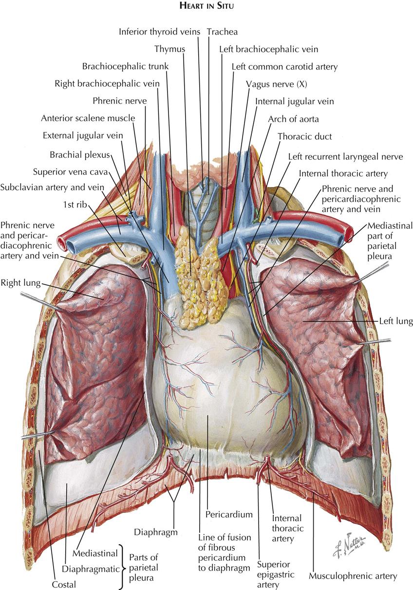
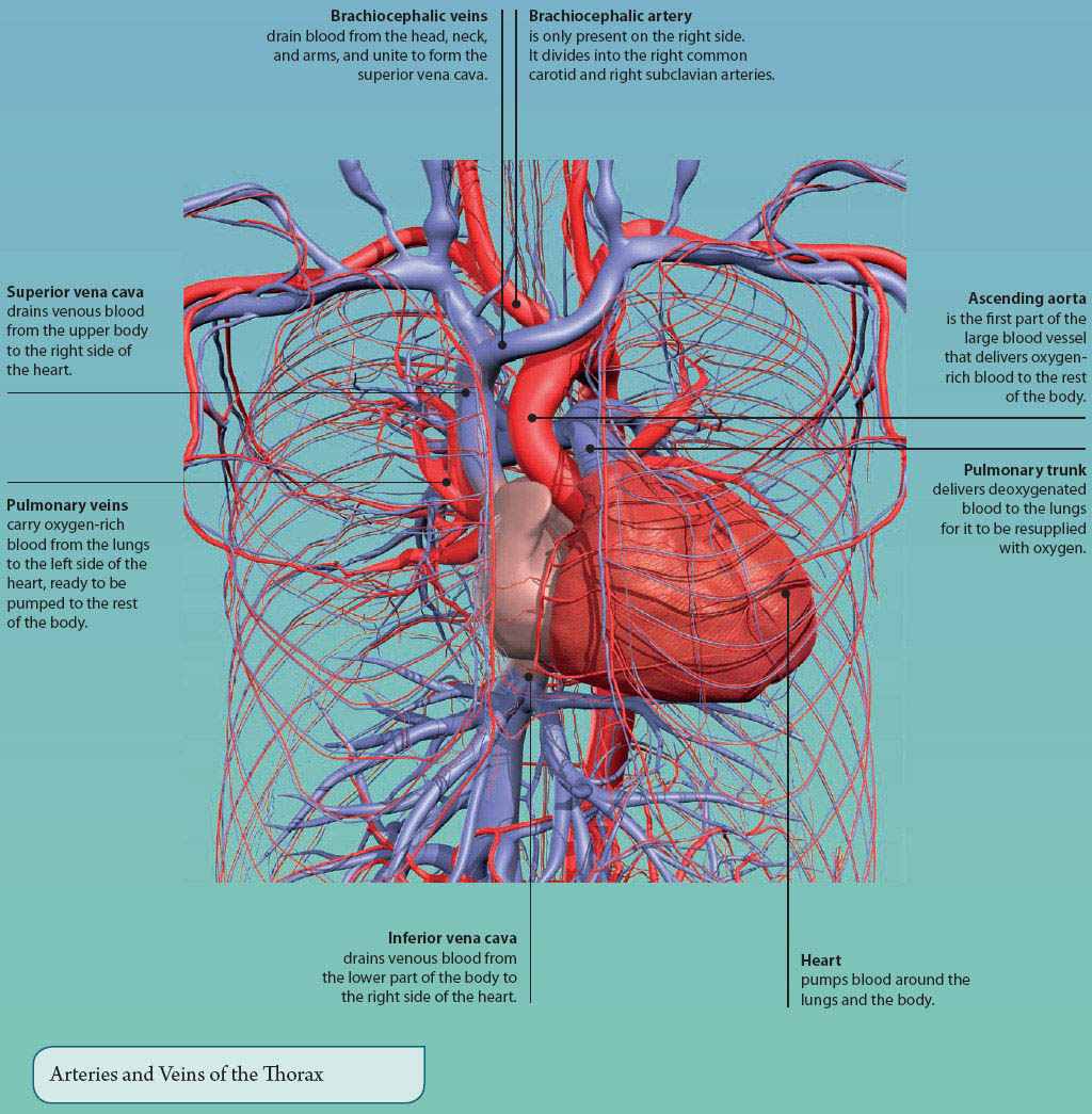
4: THE THORAX | Basicmedical Key from basicmedicalkey.com Anatomy of the thorax, heart, abdomen and pelvis recommended text gray's anatomy for students, richard l drake, elsevier. The chest, abdomen, and back elmar peuker, mike cummings. We hope you will use this picture in the study and helping your research. If you've been following some of our other lessons on muscle anatomy, you may recall that, just like the skeleton, the muscles of the body can be divided into two groups based on location. Anatomical illustrations this e anatomy module presents an illustrated anatomy of the lungs trachea bronchi pleural cavity and pulmonary vessels. The four anatomical regions of the abdomen are known as quadrants. Specialized myocytes contribute to generate electrical impulses it is subject to regulation of the autonomic nervous system by the interatrial septum crossings leading crossing system atrioventricular it is supplied by coronary arteries if ischemia may. This post human thorax and abdomen organs anatomy belong to following category/categories, you may also find more related.
About the 6th week, the somites differentiate into the sclerotomes and the dermatomyotomes.
Use the mouse scroll wheel to move the images up and down alternatively use the tiny arrows (>>) on both side of the image to move the images. Abdominal computed tomography (ct) is a type of medical imaging procedure used to diagnose and monitor internal stomach issues, like cancer, bowel obstruction, and abdominal this photo gallery presents the anatomy of the abdomen by means of ct (axial, coronal, and sagittal reconstructions). Anatomy for the acupuncturist facts & fiction. The trachea (windpipe) conducts inhaled air into the lungs through its tubular branches, called bronchi. Although chest and abdominal radiography both involve soft tissue and organs, they are quite different. Anatomy of the thorax, heart, abdomen and pelvis recommended text gray's anatomy for students, richard l drake, elsevier. Anatomy is the amazing science. Start your review of anatomy of the chest, abdomen, and pelvis. Learn about its function, parts, abdominal conditions, and more. The diaphragm forms the upper surface of. But with the use of smart technology, you can learn faster and master abdomen anatomy in no time! This post human thorax and abdomen organs anatomy belong to following category/categories, you may also find more related. Specialized myocytes contribute to generate electrical impulses it is subject to regulation of the autonomic nervous system by the interatrial septum crossings leading crossing system atrioventricular it is supplied by coronary arteries if ischemia may.
Basic information on the anatomy, positioning, and pathology of the abdomen is included here so that the content will be comprehensive for those who need this information. Of sectional anatomy, computed tomography and magnetic resonance imaging: The muscles of the chest develop from the somites found in the mesoderm. If you want to learn how to read ct scans of the abdomen and pelvis proficiently, this video is an excellent starting point. With anatomical models and charts, study of the chest and abdomen becomes a snap!
Muscles of Head, Neck, Chest, Back, Abdomen, and Shoulder ... from classconnection.s3.amazonaws.com Start your review of anatomy of the chest, abdomen, and pelvis. Find out more about the individual muscles within the chest anatomy by clicking their respective links throughout this page. The chest, abdomen, and back elmar peuker, mike cummings. The diaphragm forms the upper surface of. The four anatomical regions of the abdomen are known as quadrants. Anatomy of the chest and the lungs: Learn about the heart, lungs, intestines, and other major organs of the chest and abdomen. They are separated by theoretical anatomical lines that can be traced on the right and left hypochondriac regions are found superiorly on either side of the abdomen, while the epigastric region sits between them in a central.
The muscles of the chest develop from the somites found in the mesoderm.
Find out more about the individual muscles within the chest anatomy by clicking their respective links throughout this page. Its origin is from the lower 8 ribs, and its insertion is along the. Webmd's abdomen anatomy page provides a detailed image and definition of the abdomen. Senior lecturer anatomy knowledge, and the skill to apply it, is arguably the most important facet of safe and competent department of anatomy clinical anatomy acupuncture practice. Anatomy for the acupuncturist facts & fiction. About the 6th week, the somites differentiate into the sclerotomes and the dermatomyotomes. Although chest and abdominal radiography both involve soft tissue and organs, they are quite different. We hope you will use this picture in the study and helping your research. The four anatomical regions of the abdomen are known as quadrants. Learn about chest and abdomen anatomy with free interactive flashcards. Sciency root words make anatomical parts harder to memorize. Start your review of anatomy of the chest, abdomen, and pelvis. This mri abdomen axial cross sectional anatomy tool is absolutely free to use.
The abdomen is the part of the body that contains all of the structures between the thorax (chest) and the pelvis, and is separated from the thorax via the diaphragm anatomy of chest. Start your review of anatomy of the chest, abdomen, and pelvis.

0 Comments: